Fetal Pig Dissection
Laboratory 1, Anatomy and Physiology
Abstract. The goal was to learn the basic anatomy of the human body. However, it is not a realistic option to dissect the human body. Thus, a dissection of a fetal pig was conducted because the anatomy of a fetal pig is very similar to that of a human. After the dissection, much insight was gained on the knowledge of both the fetal pig and human body. It will provide valuable assistance in regards to the diagnosis of Grandma Tilly who died several years ago.
Introduction
Anatomy can be defined as the study of the structure of the human body. Physiology is the study of the bodily functions of living organisms. Anatomy like any other science requires multiple sources of evidence in order for something to be declared as "true." Furthermore, like any other science anatomy benefits from collaboration. Collaboration allows one to get another mind to get a look on an idea and provides a fresh point of view. Without a second scientist to provide another opinion there is no one to prove a theory wrong when it is incorrect and the wrong ideas will be taken as fact. However, what is taken as fact is subject to change if new evidence presents itself. The study of anatomy dates back as far 1600 BC in Ancient Egypt with the Edwin Smith Surgical Papyrus. The Edwin Smith Surgical Papyrus was a document, which detailed 48 cases of injuries. The document recognizes, the heart, liver, kidneys, hypothalamus, uterus, and bladder. It was discovered that the heart supplied the blood to the rest of the body via veins. The Egyptians were the first to discover the function of the lymphatic system. The functions of kidneys were not yet known. Ancient Greek scientists made more progress in this field. The scientist Alcmaeon made many discoveries in anatomical science from his dissections of animals. King Ptolemy I Soter was the first to allow the use of dead bodies for the medical purposes for discovering the operations of the human body. Most of the cadavers used were the bodies of executed criminals. In the second century, Galen uncovered many truths of anatomy through vivisection. Vivisection is surgery conducted on a living animal. Furthermore, Galen served as a doctor for gladiators. Through this Galen was able to make observations without conducting any experiments on humans.
During the Renaissance period, Leonardo da Vinci studied anatomy by observing and dissecting over 30 cadavers. Hospitals in Italy permitted him to study the corpses of patients. He displayed his anatomical knowledge in his artwork of the skeleton, skin, muscles, and other visible features. However, the Pope halted his studies. In the 17th century, Andreas Vesalius utilized the invention of the printing press and published the first essays on anatomy. Vesalius breathed new life into the field of anatomy. He demonstrated the inaccuracies in the works of previous scientists theories on anatomy. In the past before human bodies were available to be observed what was true for apes and other animals was thought to be true for humans as well. Vesalius disproved such beliefs. Vesalius then decided to examine corpses for himself and build new theories with evidence that he has gained. After learning of the ways past anatomists learned of anatomy, the decision to dissect a fetal pig was made in order to gain knowledge of the body seeing as it will not be possible to dissect the human body. The goal is to acquire valuable insight on the structure of the body. We hypothesize that the fetal pig will provide an adequate outline of the structure of the human anatomy.
Methods
This study was conducted at New Tech High @ Coppell on September 15, 2014. Our experiment was located in Mrs. Wootton’s room. We dissected fetal pigs that were given to us by Mr. Bess. The work was divided evenly between the four of us group members. Each member had their own specific role that they fulfilled.
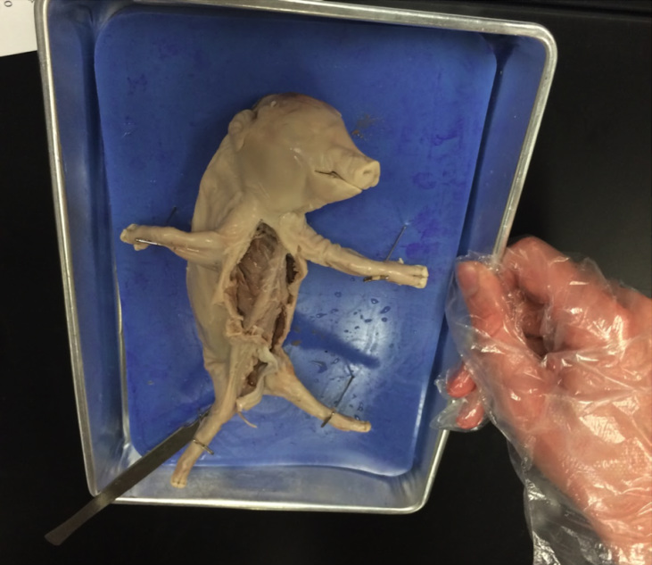
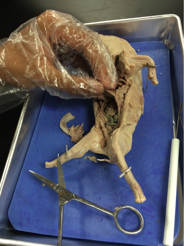
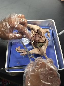
We recorded and photographed the process of us cutting open the subject and observing its insides. To begin we opened the mouth and observed the oral cavity. Afterwards, we made an incision on the ventral side of the body from below the umbilical cord all the way up to the jaw. We then opened the skin and placed pins to keep the fetal pig in place. We observed the thoracic and abdominal cavities and the organs inside. After observing what we could we used scissors to open the rib cage of the animal and allow a better view of the thoracic cavity. We freely moved organs to allow for a better view of certain tissues and organs.
Results
During the experiment we were able to gain practical knowledge of the structure of the fetal pig. The oral cavity contains the mouth, tongue, teeth, and hard and soft palate. The hard palate is formed the way it is to direct food to the rear end of the mouth. Then afterwards the soft palate can direct the food down one’s throat. The hard palate uses things such as the tongue to help so it need to be hard to allow for the pressure.
The thoracic cavity includes the rib cage, heart, thymus, trachea, bronchi, esophagus, lungs, and diaphragm. The esophagus carries food from the mouth to the digestive organs in the abdominal cavity. The trachea, also commonly referred to as windpipe, carries air from the mouth to the bronchi which transfers the air to the lungs. The lungs are aided by the muscle, diaphragm, which belongs in the respiratory and muscular systems. The lungs and heart take up the majority of the space in the thoracic cavity. The thymus is a gland located between the lungs that aids the body’s immune system. The heart has four chambers which is the same as the number of chambers in the human heart.
The abdominal cavity consists of the mesentery, adrenal glands, inferior vena cava, abdominal aorta, liver, stomach, spleen, pancreas, small and large intestine, kidneys, and urinary bladder. The liver and intestines take up the largest amount of mass in the abdominal cavity. The stomach is inferior to the liver. The intestines are coiled to allow them to be as long as possible. This allows food to be in the body for the maximum amount of time allowing for a greater period for the body to absorb nutrients.The small intestines are about 6 ft whereas the large intestines are about 1 ft. The large intestines have a much larger diameter than the small intestines. Mesentery is located posterior to the intestines and is a membrane that exists to keep the intestines in place. There is adipose (fat) tissue located around the kidney to protect the it from injury. The kidneys connect to the urinary bladder via the 2 ureters. The bladder is very difficult to locate because it is the utmost inferior organ. The adrenal gland is part of the endocrine system and produces the hormone adrenaline. The spleen is inferior to the stomach and the pancreas is the most posterior organ in the abdominal cavity. Though the inferior vena cava and the abdominal aorta are in the abdominal cavity away from the heart they belong to the cardiovascular.
Discussion
Although we have now acquired keen insight towards the inner working of the fetal pig body and there are several similarities between the pig’s body and the human body we still have not gained exact anatomical knowledge of the human body. In order to better our anatomical and physiological understanding of the human body further studies must be conducted towards human anatomy. However, this lab did allow us practical knowledge of anatomy and gave us the opportunity to carefully and closely observe the body of an animal.
Conclusion
The purpose of the dissection was to gain basic knowledge on the anatomy and structure of the fetal pig. The similarities between the human and fetal pig bodies will aid us in discovering the ailment with Grandma Tilly. We know the anatomy of the thoracic and abdominal cavities as well as the oral cavity. We are able to locate and identify various organs and discuss their functions and organ systems.
Works Cited
"Historical Anatomies on the Web: Browse Titles." U.S National Library of Medicine. U.S. National Library of Medicine, 08 June 2012. Web. 09 Sept. 2014.
"History of Anatomy." Princeton University. Princeton University, n.d. Web. 09 Sept. 2014.
Laboratory 1, Anatomy and Physiology
Abstract. The goal was to learn the basic anatomy of the human body. However, it is not a realistic option to dissect the human body. Thus, a dissection of a fetal pig was conducted because the anatomy of a fetal pig is very similar to that of a human. After the dissection, much insight was gained on the knowledge of both the fetal pig and human body. It will provide valuable assistance in regards to the diagnosis of Grandma Tilly who died several years ago.
Introduction
Anatomy can be defined as the study of the structure of the human body. Physiology is the study of the bodily functions of living organisms. Anatomy like any other science requires multiple sources of evidence in order for something to be declared as "true." Furthermore, like any other science anatomy benefits from collaboration. Collaboration allows one to get another mind to get a look on an idea and provides a fresh point of view. Without a second scientist to provide another opinion there is no one to prove a theory wrong when it is incorrect and the wrong ideas will be taken as fact. However, what is taken as fact is subject to change if new evidence presents itself. The study of anatomy dates back as far 1600 BC in Ancient Egypt with the Edwin Smith Surgical Papyrus. The Edwin Smith Surgical Papyrus was a document, which detailed 48 cases of injuries. The document recognizes, the heart, liver, kidneys, hypothalamus, uterus, and bladder. It was discovered that the heart supplied the blood to the rest of the body via veins. The Egyptians were the first to discover the function of the lymphatic system. The functions of kidneys were not yet known. Ancient Greek scientists made more progress in this field. The scientist Alcmaeon made many discoveries in anatomical science from his dissections of animals. King Ptolemy I Soter was the first to allow the use of dead bodies for the medical purposes for discovering the operations of the human body. Most of the cadavers used were the bodies of executed criminals. In the second century, Galen uncovered many truths of anatomy through vivisection. Vivisection is surgery conducted on a living animal. Furthermore, Galen served as a doctor for gladiators. Through this Galen was able to make observations without conducting any experiments on humans.
During the Renaissance period, Leonardo da Vinci studied anatomy by observing and dissecting over 30 cadavers. Hospitals in Italy permitted him to study the corpses of patients. He displayed his anatomical knowledge in his artwork of the skeleton, skin, muscles, and other visible features. However, the Pope halted his studies. In the 17th century, Andreas Vesalius utilized the invention of the printing press and published the first essays on anatomy. Vesalius breathed new life into the field of anatomy. He demonstrated the inaccuracies in the works of previous scientists theories on anatomy. In the past before human bodies were available to be observed what was true for apes and other animals was thought to be true for humans as well. Vesalius disproved such beliefs. Vesalius then decided to examine corpses for himself and build new theories with evidence that he has gained. After learning of the ways past anatomists learned of anatomy, the decision to dissect a fetal pig was made in order to gain knowledge of the body seeing as it will not be possible to dissect the human body. The goal is to acquire valuable insight on the structure of the body. We hypothesize that the fetal pig will provide an adequate outline of the structure of the human anatomy.
Methods
This study was conducted at New Tech High @ Coppell on September 15, 2014. Our experiment was located in Mrs. Wootton’s room. We dissected fetal pigs that were given to us by Mr. Bess. The work was divided evenly between the four of us group members. Each member had their own specific role that they fulfilled.
We recorded and photographed the process of us cutting open the subject and observing its insides. To begin we opened the mouth and observed the oral cavity. Afterwards, we made an incision on the ventral side of the body from below the umbilical cord all the way up to the jaw. We then opened the skin and placed pins to keep the fetal pig in place. We observed the thoracic and abdominal cavities and the organs inside. After observing what we could we used scissors to open the rib cage of the animal and allow a better view of the thoracic cavity. We freely moved organs to allow for a better view of certain tissues and organs.
Results
During the experiment we were able to gain practical knowledge of the structure of the fetal pig. The oral cavity contains the mouth, tongue, teeth, and hard and soft palate. The hard palate is formed the way it is to direct food to the rear end of the mouth. Then afterwards the soft palate can direct the food down one’s throat. The hard palate uses things such as the tongue to help so it need to be hard to allow for the pressure.
The thoracic cavity includes the rib cage, heart, thymus, trachea, bronchi, esophagus, lungs, and diaphragm. The esophagus carries food from the mouth to the digestive organs in the abdominal cavity. The trachea, also commonly referred to as windpipe, carries air from the mouth to the bronchi which transfers the air to the lungs. The lungs are aided by the muscle, diaphragm, which belongs in the respiratory and muscular systems. The lungs and heart take up the majority of the space in the thoracic cavity. The thymus is a gland located between the lungs that aids the body’s immune system. The heart has four chambers which is the same as the number of chambers in the human heart.
The abdominal cavity consists of the mesentery, adrenal glands, inferior vena cava, abdominal aorta, liver, stomach, spleen, pancreas, small and large intestine, kidneys, and urinary bladder. The liver and intestines take up the largest amount of mass in the abdominal cavity. The stomach is inferior to the liver. The intestines are coiled to allow them to be as long as possible. This allows food to be in the body for the maximum amount of time allowing for a greater period for the body to absorb nutrients.The small intestines are about 6 ft whereas the large intestines are about 1 ft. The large intestines have a much larger diameter than the small intestines. Mesentery is located posterior to the intestines and is a membrane that exists to keep the intestines in place. There is adipose (fat) tissue located around the kidney to protect the it from injury. The kidneys connect to the urinary bladder via the 2 ureters. The bladder is very difficult to locate because it is the utmost inferior organ. The adrenal gland is part of the endocrine system and produces the hormone adrenaline. The spleen is inferior to the stomach and the pancreas is the most posterior organ in the abdominal cavity. Though the inferior vena cava and the abdominal aorta are in the abdominal cavity away from the heart they belong to the cardiovascular.
Discussion
Although we have now acquired keen insight towards the inner working of the fetal pig body and there are several similarities between the pig’s body and the human body we still have not gained exact anatomical knowledge of the human body. In order to better our anatomical and physiological understanding of the human body further studies must be conducted towards human anatomy. However, this lab did allow us practical knowledge of anatomy and gave us the opportunity to carefully and closely observe the body of an animal.
Conclusion
The purpose of the dissection was to gain basic knowledge on the anatomy and structure of the fetal pig. The similarities between the human and fetal pig bodies will aid us in discovering the ailment with Grandma Tilly. We know the anatomy of the thoracic and abdominal cavities as well as the oral cavity. We are able to locate and identify various organs and discuss their functions and organ systems.
Works Cited
"Historical Anatomies on the Web: Browse Titles." U.S National Library of Medicine. U.S. National Library of Medicine, 08 June 2012. Web. 09 Sept. 2014.
"History of Anatomy." Princeton University. Princeton University, n.d. Web. 09 Sept. 2014.